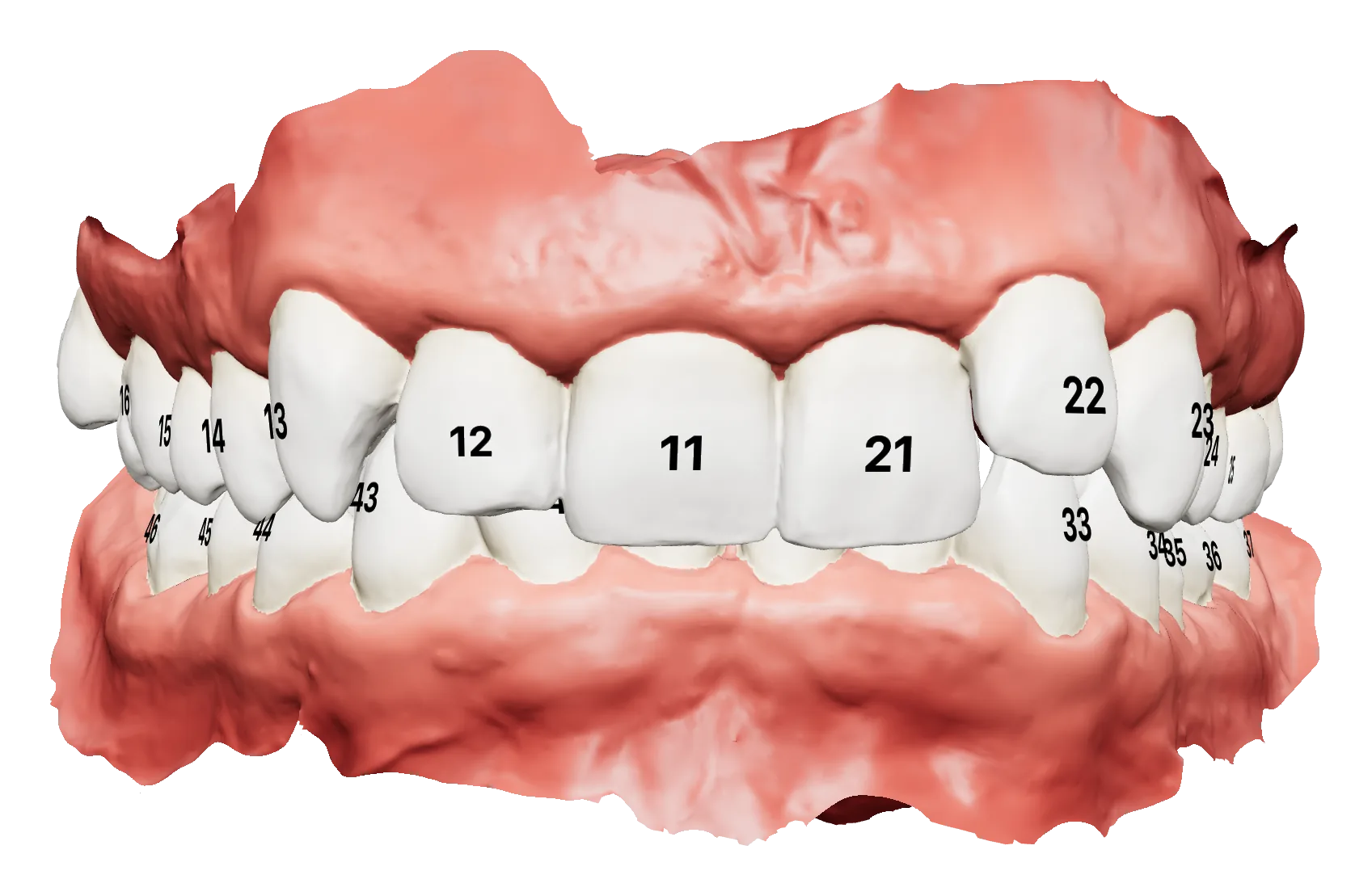
Every tooth, segmented and numbered
Upload the upper and lower scans: each tooth is separated from the gingiva and given its FDI number, permanent and deciduous alike.
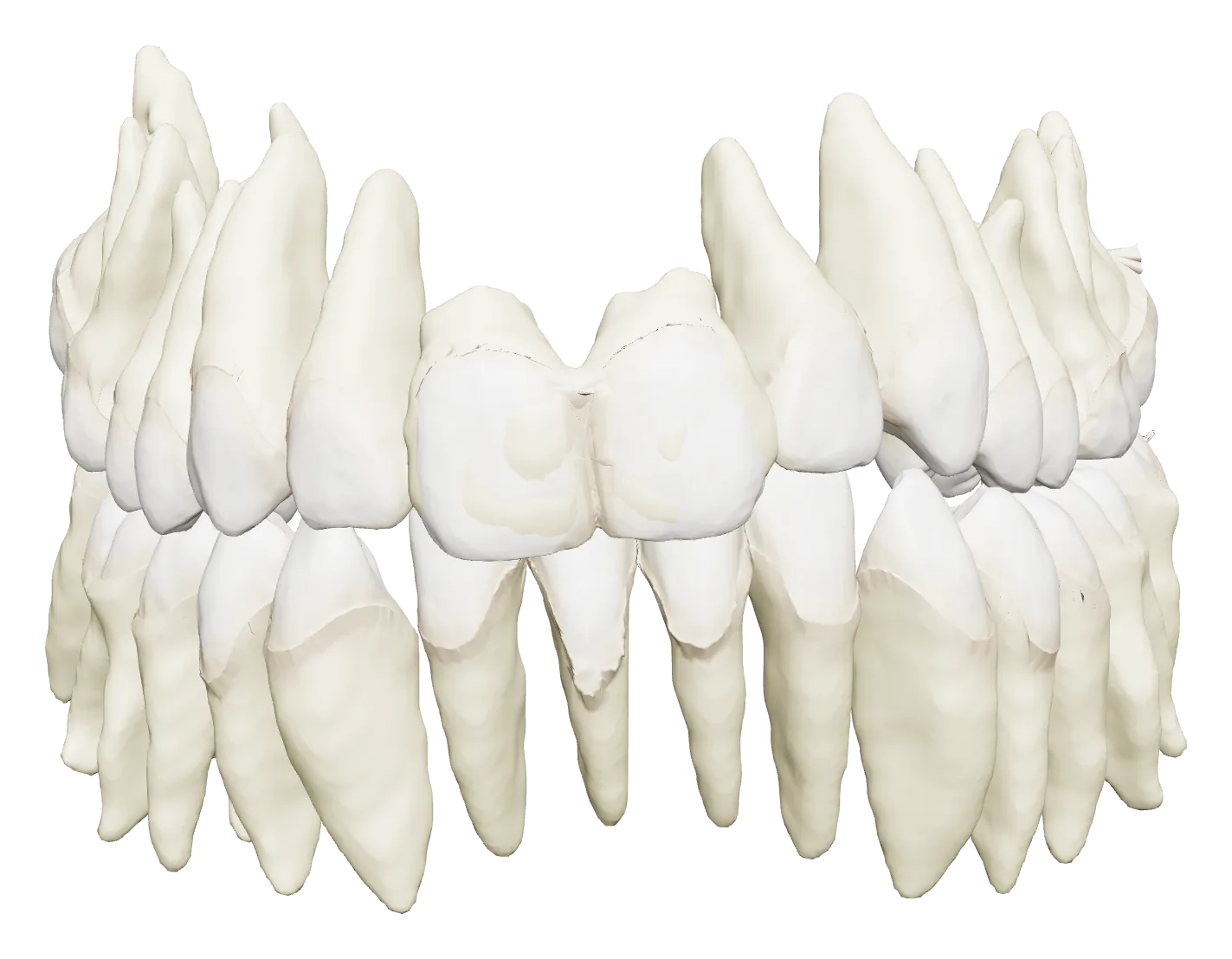
The real roots, from the CBCT
Load the patient's CBCT: teeth and roots are segmented and fused with the scanned crowns, so every movement is planned on the whole tooth.
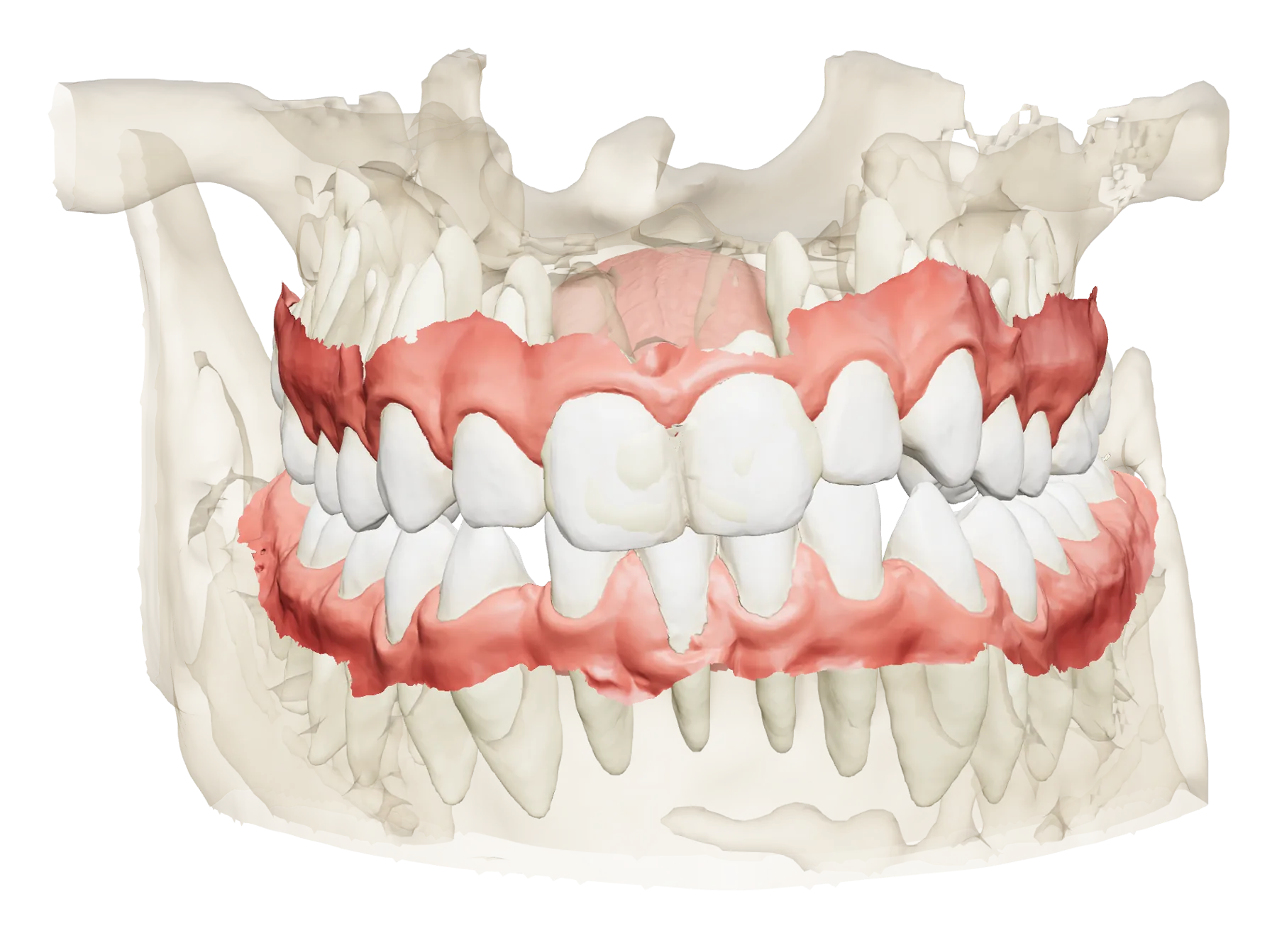
Scans and bone in one view
The intraoral scans are superimposed on the CBCT, with the bone around them, to see where the teeth can move.
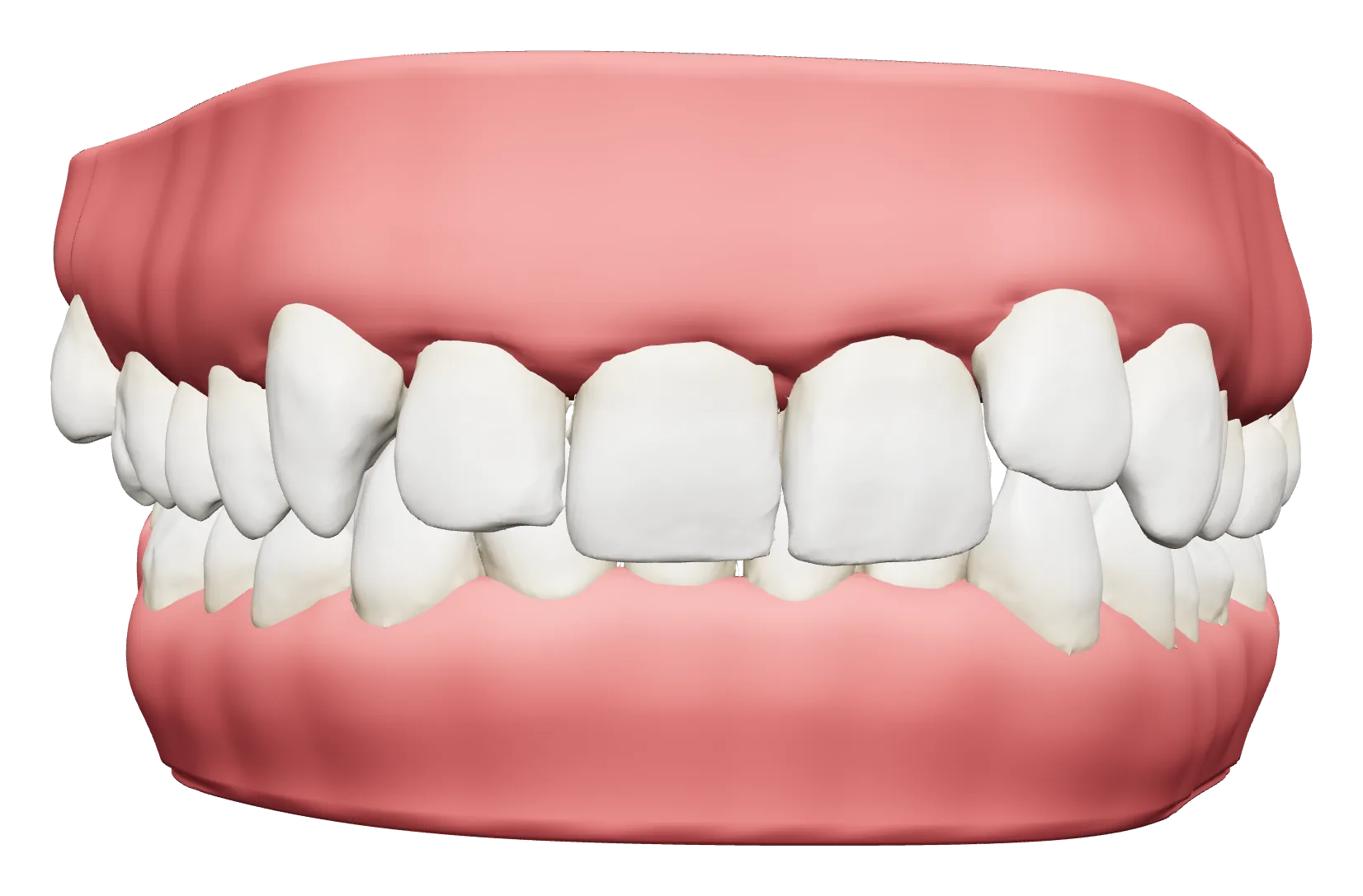
Plan and stage the treatment
Move each tooth in six directions, stage the movements, compare two versions side by side and export a printable model for every step.
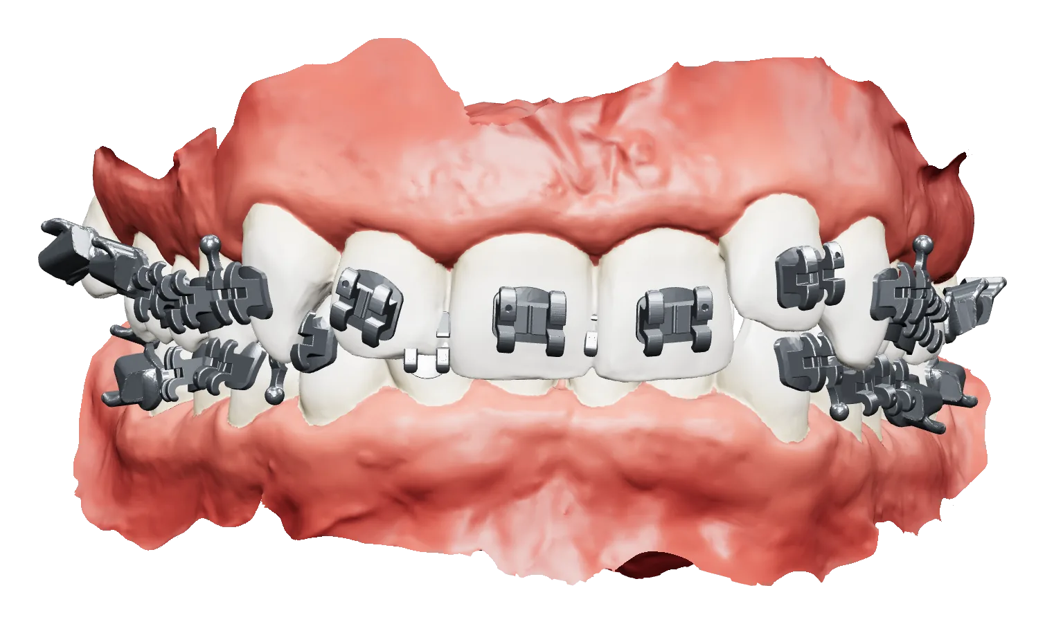
Brackets placed for you
Brackets are positioned at the FA point or from a positioning chart, then adjusted tooth by tooth, ready for indirect bonding.
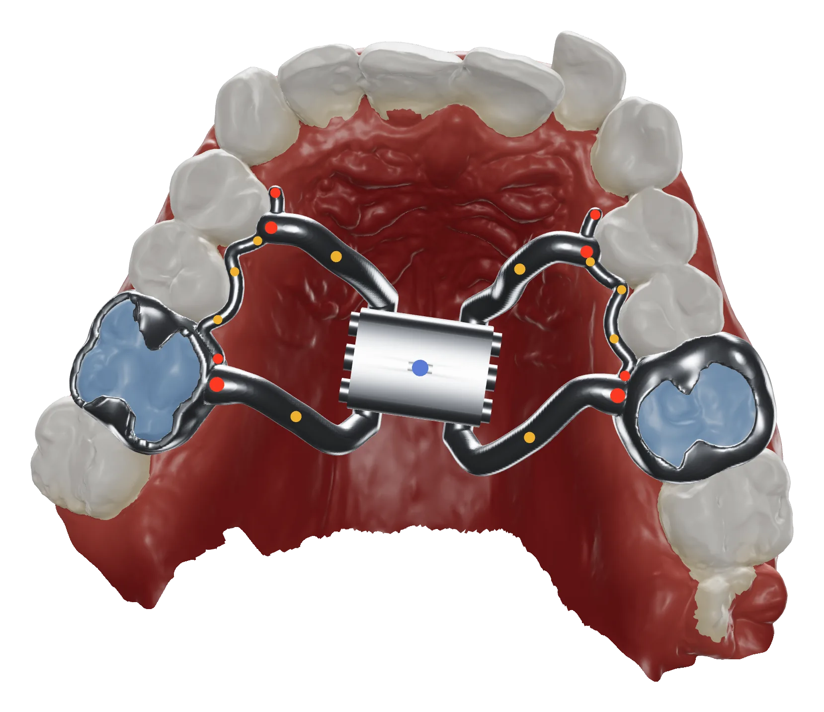
Bands and expanders, designed on the arch
Bands on the teeth you choose and a rapid palatal expander shaped on the patient's palate, exported for printing.
From the scan to the appliance
One web application for the whole digital workflow, with nothing to install.
Segmentation and numbering
Teeth and gingiva separated automatically on upper and lower scans, with FDI numbering.
CBCT fusion
Roots and bone from the CBCT, superimposed on the scans and exported as STL.
Treatment simulator
Movements in six directions, advanced staging, collisions, IPR, pontics and two versions to compare.
Printable steps
A model for every step of the treatment, with closed gingiva, ready for the printer.
Indirect bonding
Brackets positioned automatically and refined tooth by tooth.
Bands and expanders
Bands and rapid palatal expanders designed on the patient's arch.
Photos and radiographs
Patient photos and radiographs organised in one place, with automatic analysis and a PDF report.
Cephalometric tracing
Landmarks traced on the lateral cephalogram, ready to be checked and corrected.
Request information
Tell us about your practice or laboratory and we will get back to you about access, a demonstration or a trial.
- Works in the browser, nothing to install
- Accounts for clinicians and laboratories
- Developed by the Ferrara School of Orthodontics
Already have an account? Log in